Background Questions
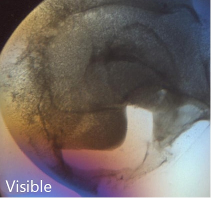
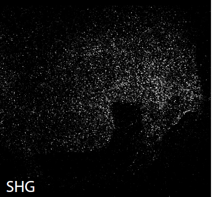
Are all protein crystals detectable?
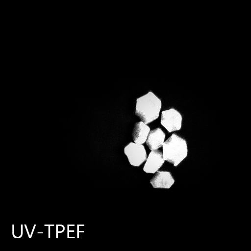
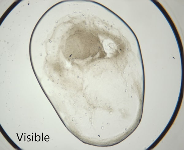
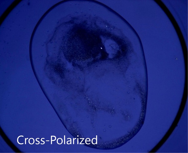
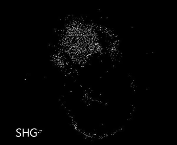
Almost all molecules that have a chiral center form a chiral crystal. Therefore, most proteins will form chiral crystals that are detectable via SONICC® (Second Order Non-linear Imaging of Chiral Crystals). Over 99% of the proteins in the Protein Data Bank have a space group that is detectable with SONICC. Crystals with extremely high symmetry classes will generate less SHG signal. Although theoretically all protein crystals should be detected with SHG, that is not always experimentally the case. The higher the space group, the less SHG is generated. Also, the hyperpolarizability (ie the SHG efficiency) of the protein significantly impacts the amount of light generated. Some proteins interact more favorably with light and therefore produce higher SHG signal, where other proteins hold their electrons closely, have a low hyperpolarizability and not give a strong, detectable SHG response.
What is SHG?
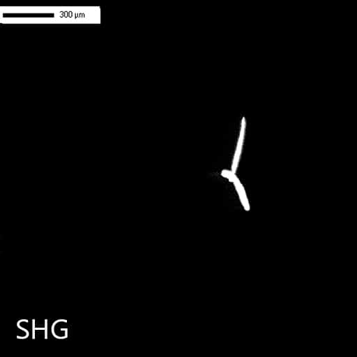
SHG stands for Second Harmonic Generation and is a nonlinear optical process. In intense electric fields (i.e. in the presence of a femtosecond laser) the distance between the electrons and the nucleus are distorted (anharmonicity) resulting in non-linear optical effects such as SHG where the frequency of the outgoing light is twice that of the incident (i.e. 1064 nm incident results in 532 nm exiting).
What does "chiral" mean?
A chiral molecule, or in this case a chiral crystal, is a crystal that lacks an internal plane of symmetry, and thus its mirror image is nonsuperimposable. Achiral crystals are symmetric and therefore produce SHG in equal and opposite directions that sum to a net zero signal.
Will salts produce signal?
They can if they are chiral, but the majority of salts are achiral and therefore do not generate SHG signal.
How is SONICC different than fluorescent imaging?
Fluorescent imaging takes advantage of either the endogenous fluorescence of the protein or the use of fluorescent tags. Although fluorescence is bright and easily detectable, it is generated from solubilized and aggregated proteins as well as crystallized proteins. The background from the solubilized protein decreases the signal to noise ratio significantly, resulting in false positives. SONICC, on the other hand, is only sensitive to crystallized proteins.
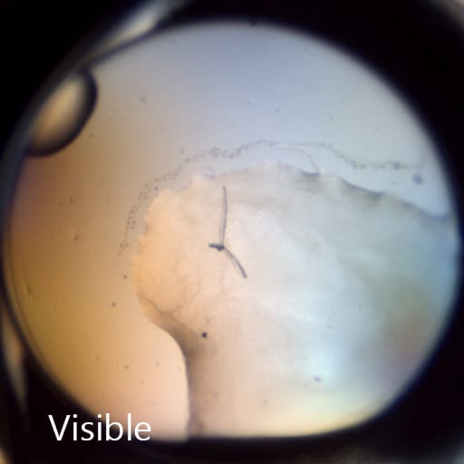
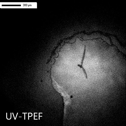
How does UV-TPEF compare to traditional UV imaging?
Both imaging methods probe the amino acids present in proteins that are excited by UV light (~280 nm). However, with UV-TPEF, the incident wavelength is 532 nm instead of UV and has less energy. UV imaging can cause the breakage of disulfide bonds but by using green light instead to excite, damage does not occur.
With which platforms are SONICC compatible?
SONICC is compatible with all optically-accessible plates and seals.
Will the laser damage my crystals?
Experiments show no detectable damage to protein crystals. In one experiment, a protein crystal was imaged on one half with excessive laser input. X-ray diffraction was obtained from both the exposed and un-exposed halves of the crystal. Both sides diffracted to within expected resolution (~2 Ã) and within statistical variation (i.e. there was no statistical difference between the diffraction of either side). SONICC has also imaged live cells with no observed impact (they remained adhered to a polylysine-coated slide).
Can I use SONICC if my sample is fluorescent?
Yes. As long as fluorescence is Stokes-shifted by 10 nm, the fluorescence will not be detected nor interfere with the SHG signal.
Specification Questions
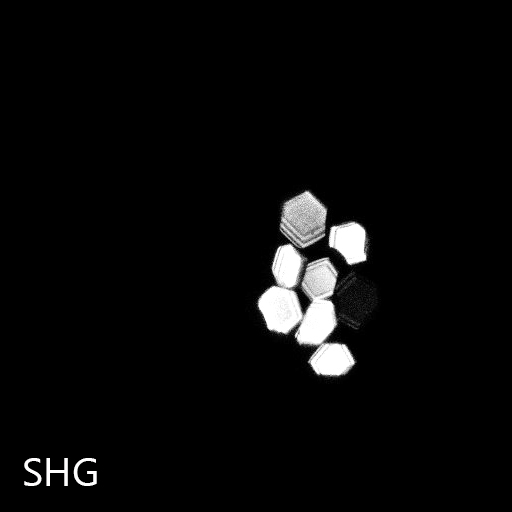
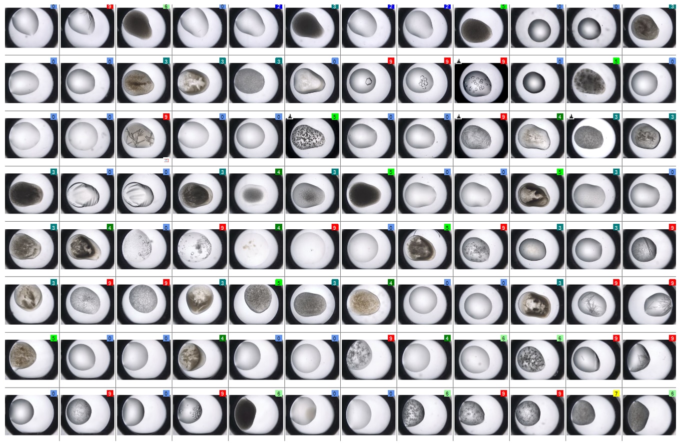
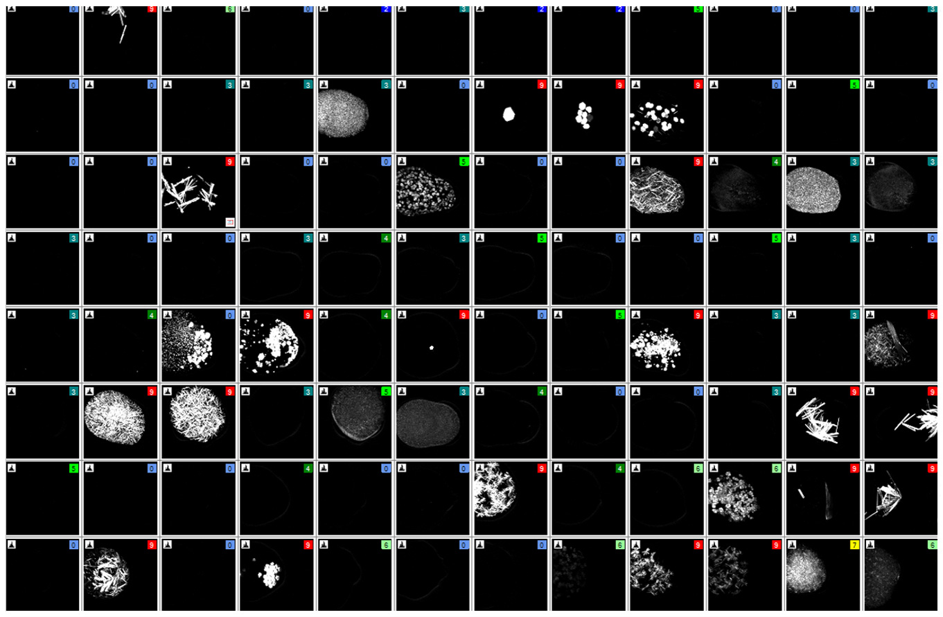
*Please note that each crystal will generate different intensities of SHG signal depending on size, orientation, space group and quality, as well as the acquisition time and incident intensity.
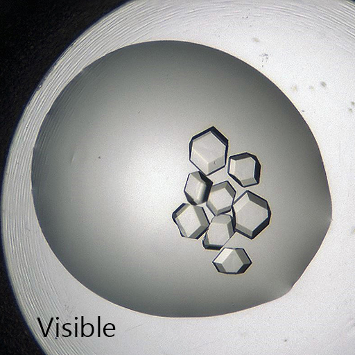
How small of a crystal can SONICC detect?
Theoretically, the lower limit of detection can be estimated by the forward-to-backward ratio of the SHG signal. Based on the coherence length of the generated SHG signal and the refractive index of the material, this lower limit ranges from 90 nm - 300 nm in thickness. In practice, 1 μm^3 crystals can be routinely detected. 2D crystals have also been routinely imaged with a signal to noise ratio >30.
What is the spatial resolution?
Crystals can be resolved to 2 μm (detection limit is much lower).
What is the laser´s Z-resolution and how deeply can it penetrate?
The laser focuses to a width of ∼50 μm and can image drops >3 mm tall with multiple Z-steps, called "slices".
How fast is SONICC?
The current electronic package allows 512 x 512 image acquisition for one Z-slice in 500 ms. This corresponds to eight traces of the fast-scanning mirror per line. A one-drop 96-well plate can be imaged with visible light in 3 minutes and with SHG in 15 minutes with eight Z-slices per drop.


")


")
")